Craniotomy For Hematoma Evacuation
Craniotomy for hematoma evacuation is brain surgery that removes a blood clot to reduce pressure on the brain when appropriate.
Craniotomy for brain tumor resection is brain surgery that creates a temporary opening in the skull so a neurosurgeon can reach and remove a brain tumor. “Craniotomy” means opening the skull. “Resection” means surgically removing tissue.
The goal is not simply to remove tumor at all costs. The goal is to remove as much tumor as safely appropriate while protecting important brain functions such as movement, speech, vision, memory, coordination, and sensation.
Brain tumor surgery may also help obtain tissue for diagnosis. A pathologist examines the tissue to determine the tumor type, grade, molecular features when appropriate, and whether additional treatment such as radiation, chemotherapy, targeted therapy, or observation may be needed.
De Novo Brain & Spine evaluates adult patients in Stockbridge, Fayetteville, Atlanta, and surrounding communities to help determine whether craniotomy for brain tumor resection, biopsy, observation, radiation, medical oncology, or another treatment plan may be appropriate.
Craniotomy for brain tumor resection may be considered for selected tumors or masses in the brain. It is not appropriate for every brain tumor, and some tumors are better treated with biopsy, radiation, chemotherapy, radiosurgery, surveillance, or multidisciplinary care.
Conditions or situations that may lead to consideration of craniotomy for tumor resection include:
The treatment decision depends on tumor location, tumor type, size, growth pattern, symptoms, neurologic function, overall health, and whether the tumor can be safely approached.
Craniotomy for brain tumor resection may be considered when a tumor is causing symptoms, growing over time, pressing on nearby brain structures, producing swelling, or creating a need for tissue diagnosis.
Symptoms that may lead to evaluation include seizures, headaches, weakness, numbness, balance problems, confusion, personality or behavior changes, speech difficulty, vision changes, memory problems, or worsening neurologic function. Some brain tumors are found incidentally on imaging done for another reason.
Surgery may be discussed when removing tumor tissue could reduce pressure, improve diagnostic certainty, help guide oncology treatment, or address symptoms caused by mass effect. In some cases, surgery is urgent because the tumor is causing rapid neurologic decline, severe swelling, hydrocephalus, hemorrhage, or dangerous pressure inside the skull.
Some tumors cannot be safely removed completely because they involve or sit near critical brain areas. In those situations, the surgical goal may be partial removal, biopsy, decompression, or referral for additional treatment rather than complete resection.
Doctors determine whether craniotomy for brain tumor resection may be appropriate by reviewing the patient’s symptoms, neurologic examination, imaging, tumor location, medical history, and treatment goals.
Evaluation may include:
The decision depends on whether surgery is likely to provide useful diagnosis, reduce pressure, improve safety of further treatment, or remove tumor without unacceptable risk to brain function.
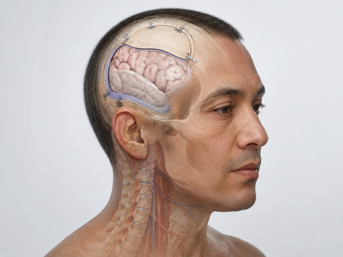
Craniotomy for brain tumor resection is performed under anesthesia. The surgeon makes an incision in the scalp and temporarily removes a section of skull bone to reach the area of the tumor.
The covering of the brain, called the dura, is opened. The surgeon then uses imaging, anatomy, the operating microscope, surgical instruments, and sometimes computer-assisted navigation or brain mapping to locate and remove tumor tissue as safely as possible.
The amount of tumor removed depends on the tumor type, borders, location, relationship to critical brain structures, and how safely the tumor can be separated from normal brain tissue. Some tumors are well-defined, while others grow into surrounding brain tissue and cannot be fully separated.
After tumor removal, the dura is closed, the bone flap is usually replaced and secured, and the scalp is closed. Tissue removed during surgery is sent to pathology for diagnosis.
The main goals of craniotomy for brain tumor resection are to remove as much tumor as safely appropriate, obtain tissue for diagnosis, reduce pressure on nearby brain structures, and support additional treatment planning.
Potential benefits may include reduction of mass effect, improved diagnostic clarity, seizure control in selected cases, relief of pressure-related symptoms, or improved ability to plan radiation, chemotherapy, targeted therapy, or observation. Improvement is not guaranteed.
Craniotomy for tumor resection has important limitations. It may not cure the tumor. It may not remove all tumor cells, especially when the tumor grows into surrounding brain tissue. It does not guarantee neurologic recovery, seizure control, tumor control, or survival benefit for every patient.
General risks may include infection, bleeding, seizure, stroke, brain swelling, spinal fluid leak, weakness, numbness, speech difficulty, vision changes, memory or thinking changes, personality changes, wound problems, blood clots, need for additional surgery, or other complications. Risks depend on tumor location, tumor type, brain function near the tumor, patient health, and the surgical plan.
Treatment planning for brain tumors is individualized and often multidisciplinary. Craniotomy for tumor resection is one possible treatment option.
Other options may include:
These treatments are not interchangeable. The best plan depends on tumor type, tumor location, symptoms, growth rate, neurologic function, overall health, patient goals, and whether tissue diagnosis has already been established.
Recovery after craniotomy for brain tumor resection varies from person to person. It depends on tumor location, tumor type, extent of surgery, neurologic status before surgery, swelling, seizure history, overall health, and whether additional treatment is needed.
Follow-up usually focuses on incision healing, neurologic function, headaches, seizure monitoring, medication management, pathology results, and postoperative imaging. Patients may need additional care with radiation oncology, medical oncology, neuro-oncology, neurology, rehabilitation, or primary care.
Pathology results are especially important. The diagnosis may guide whether the next step is observation, radiation, chemotherapy, targeted treatment, clinical trial discussion, or additional surgery.
Some symptoms may improve after surgery, while others may persist or recover slowly. In some cases, new neurologic symptoms may occur after surgery and may require rehabilitation or additional treatment.
Seek emergency medical care or call 911 for a new seizure, sudden weakness, numbness on one side of the body, trouble speaking, confusion, loss of consciousness, sudden severe headache, rapid neurologic decline, or new vision loss.
Urgent evaluation is also important for worsening headaches with vomiting, severe sleepiness, new balance problems, sudden personality changes, fever with worsening headache, or symptoms that suggest increased pressure inside the skull.
After brain tumor surgery, patients should seek urgent medical care for fever, worsening incision redness or drainage, severe headache, new seizure, worsening confusion, weakness, speech difficulty, fluid leakage from the incision, or any neurologic symptom that is new or rapidly worsening.
Craniotomy for brain tumor resection is used to remove as much tumor as safely appropriate, obtain tissue for diagnosis, reduce pressure on nearby brain structures, and help guide additional treatment planning.
Sometimes, but not always. The amount of tumor that can be removed depends on the tumor type, location, borders, and relationship to critical brain areas. In some cases, partial removal or biopsy is safer than complete removal.
No. Some tumors are treated first with observation, biopsy, radiation, radiosurgery, chemotherapy, targeted therapy, or a combination of treatments. The best plan depends on the tumor type, symptoms, location, growth pattern, and the patient’s overall condition.
Tissue diagnosis allows a pathologist to identify the tumor type and grade. In many cases, pathology and molecular testing help guide decisions about radiation, chemotherapy, targeted therapy, follow-up imaging, or additional treatment.
Seek urgent medical care for a new seizure, sudden weakness, trouble speaking, confusion, sudden severe headache, loss of consciousness, new vision loss, or rapid neurologic decline. These symptoms may suggest a serious brain condition that needs prompt evaluation.
Schedule a Consultation
Learn if this procedure is right for you.
Craniotomy for hematoma evacuation is brain surgery that removes a blood clot to reduce pressure on the brain when appropriate.
Hematoma evacuation removes selected blood collections in or around the brain when bleeding causes pressure, neurologic symptoms, or urgent risk.
Cranioplasty is brain surgery that repairs or reconstructs a skull defect after injury, prior surgery, infection, or bone removal.