Recurrent Brain Tumors
Evaluation and treatment planning for recurrent brain tumors, when a previously treated brain tumor returns, grows, or progresses on follow-up imaging.
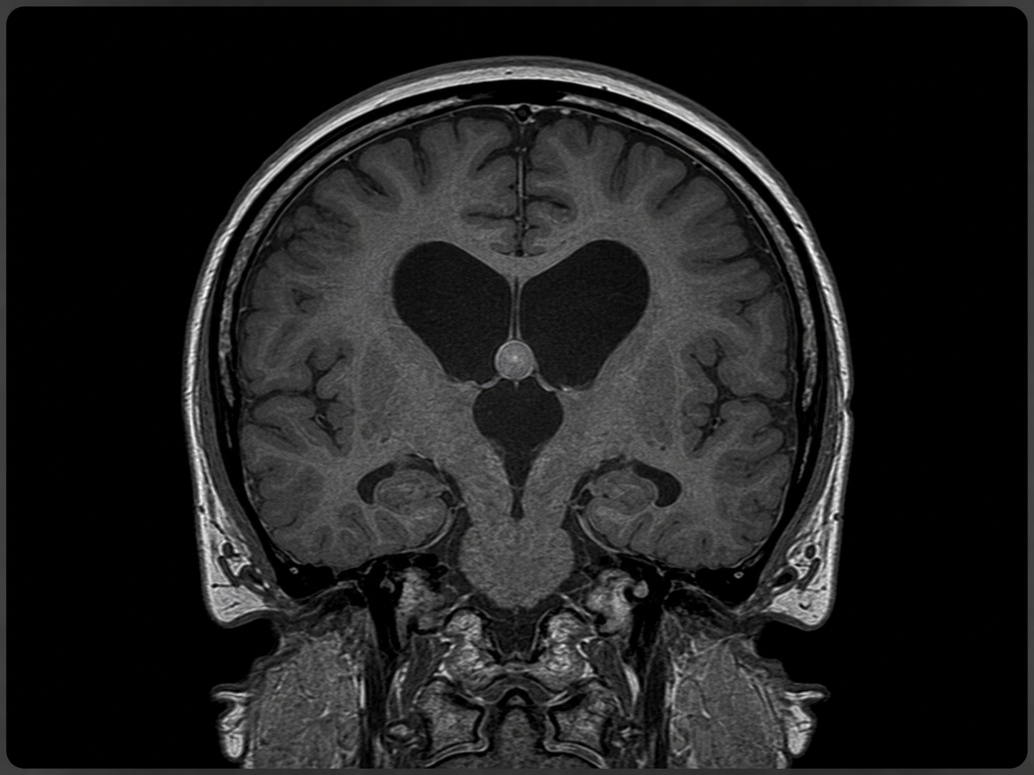
A colloid cyst is a benign cystic lesion that most often forms in the third ventricle, a fluid-filled space near the center of the brain. Colloid cysts are not cancer, but their location can be important because they may block the normal flow of cerebrospinal fluid, also called CSF.
When a colloid cyst blocks CSF flow near the foramen of Monro, fluid can build up in the brain and cause hydrocephalus. Hydrocephalus means that too much cerebrospinal fluid is collecting in the ventricles, which can increase pressure inside the head and cause neurological symptoms.
De Novo Brain & Spine evaluates adult patients with suspected or confirmed colloid cysts to help determine the appropriate next step. Evaluation may include neurological examination, brain MRI, CT imaging, review of ventricular size, monitoring with repeat imaging, or surgical consultation when symptoms or hydrocephalus are present.
Colloid cyst symptoms depend on the size of the cyst, whether it blocks cerebrospinal fluid flow, and whether hydrocephalus is present. Some colloid cysts are found incidentally on imaging and may not cause symptoms.
Common signs and symptoms may include:
Seek emergency medical care or call 911 for a sudden severe headache, repeated vomiting, fainting, new confusion, sudden weakness, seizure, loss of consciousness, or rapid neurological decline.
The exact cause of most colloid cysts is not clearly known. Many are described as developmental cysts, meaning they may form from tissue present during early development rather than from cancer or spread of disease.
Important facts about colloid cyst causes include:
The clinical concern with a colloid cyst is usually its location and effect on cerebrospinal fluid flow, not cancer behavior.
A colloid cyst cannot be diagnosed by symptoms alone. Diagnosis usually depends on medical history, neurological examination, and brain imaging.
Common diagnostic steps may include:
The goal of diagnosis is to determine whether the colloid cyst is causing symptoms, blocking cerebrospinal fluid flow, enlarging over time, or creating a risk of hydrocephalus.
Colloid cyst treatment depends on the cyst’s size, location, symptoms, ventricular size, hydrocephalus, imaging findings, neurological examination, and the patient’s overall health. Some colloid cysts can be monitored, while others may require surgery.
Treatment options may include:
Surgery may be considered when a colloid cyst is causing symptoms, obstructing cerebrospinal fluid flow, causing hydrocephalus, increasing in size, or judged to carry a higher clinical risk. The safest plan depends on the patient’s imaging, symptoms, and overall condition.
Schedule a Consultation
Get an expert opinion about your condition.
Evaluation and treatment planning for recurrent brain tumors, when a previously treated brain tumor returns, grows, or progresses on follow-up imaging.
Evaluation and treatment planning for glossopharyngeal neuralgia, a cranial nerve pain condition affecting the throat, tongue, tonsil, ear, or jaw region.
Evaluation and treatment planning for pituitary tumors that may affect hormone function, vision, headaches, and nearby skull base structures.