Craniopharyngioma

Evaluation and treatment planning for craniopharyngioma, a benign skull base tumor near the pituitary gland, optic nerves, and hypothalamus.
MRI-style image showing craniopharyngioma near the pituitary gland and optic chiasm

What is Craniopharyngioma?

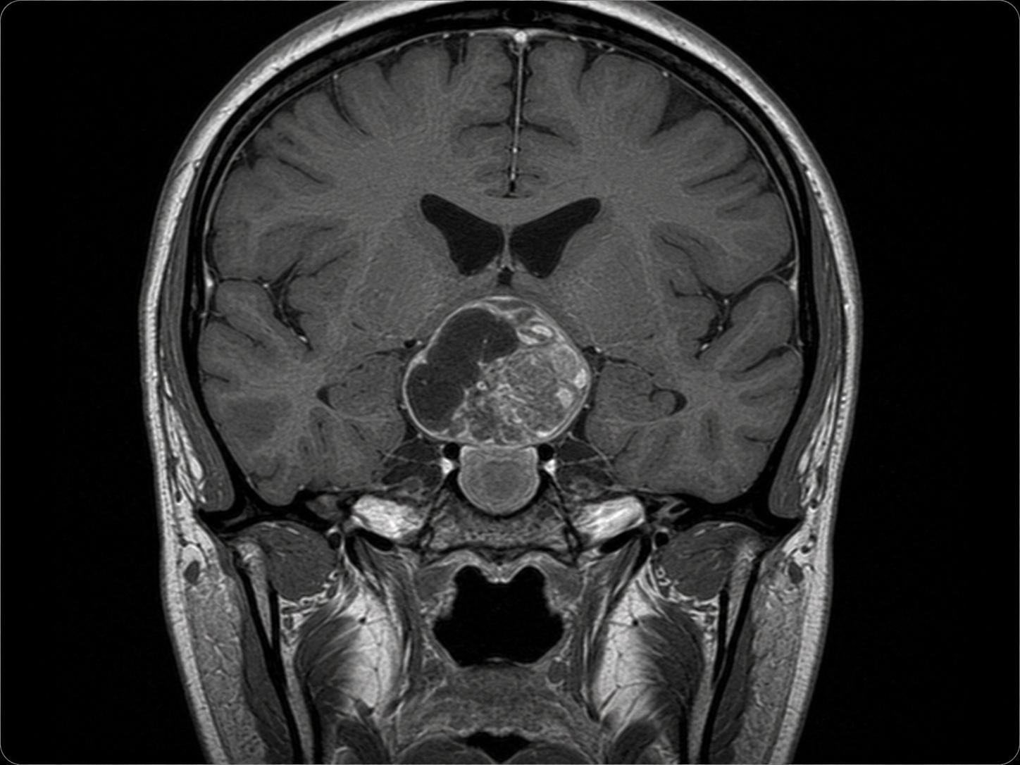
Craniopharyngioma is a usually benign brain tumor that develops near the pituitary gland at the base of the brain. It often forms in the sellar or suprasellar region, close to the optic nerves, optic chiasm, hypothalamus, and pathways that control hormone function.

Although craniopharyngiomas are not usually cancerous, their location can make them complex. A craniopharyngioma may affect vision, hormone production, memory, thirst, appetite, sleep, growth in younger patients, or the normal flow of cerebrospinal fluid, also called CSF.

De Novo Brain & Spine evaluates adult patients with suspected or confirmed craniopharyngioma to help determine the appropriate next step. Evaluation may include neurological examination, brain and pituitary MRI, CT imaging, visual testing, pituitary hormone testing, observation, surgical consultation, or coordination with endocrinology, ophthalmology, radiation oncology, or neuro-oncology when needed.

Common Signs and Symptoms

Craniopharyngioma symptoms depend on the tumor’s size, location, cystic or solid features, and effect on the optic pathways, pituitary gland, hypothalamus, or cerebrospinal fluid flow.

Common signs and symptoms may include:

  • Headaches
  • Nausea or vomiting, especially if pressure in the head increases
  • Blurred vision, double vision, or loss of peripheral vision
  • Trouble with balance or walking
  • Fatigue or low energy
  • Changes in thirst or urination
  • Unexplained weight changes or appetite changes
  • Menstrual changes, sexual dysfunction, or other hormone-related symptoms
  • Memory problems, confusion, or difficulty concentrating
  • Sleep changes or temperature regulation problems
  • Symptoms of hydrocephalus, such as worsening headache, vomiting, drowsiness, or decreased alertness

Seek emergency medical care or call 911 for sudden vision loss, severe or rapidly worsening headache, repeated vomiting, seizure, fainting, severe confusion, loss of consciousness, or rapid neurological decline.

What Causes This Condition

The exact cause of most craniopharyngiomas is not clearly known. A diagnosis of craniopharyngioma does not usually mean that the patient did something to cause the tumor.

Important facts about craniopharyngioma causes and classification include:

  • Craniopharyngiomas are usually benign, meaning noncancerous
  • They are thought to arise from tissue related to early development of the pituitary region
  • They most often occur near the pituitary gland, optic chiasm, and hypothalamus
  • They are not usually caused by head injury, routine activity, or lifestyle factors
  • There are different tumor subtypes, including adamantinomatous craniopharyngioma and papillary craniopharyngioma
  • Molecular findings, such as BRAF V600E in some papillary craniopharyngiomas, may be relevant in selected cases

These classification and molecular details are not lifestyle causes. They help doctors understand the tumor type and may help guide treatment planning.

How It Is Diagnosed

Craniopharyngioma cannot be diagnosed by symptoms alone. Diagnosis usually requires medical history, neurological examination, imaging, and evaluation of vision and hormone function.

Common diagnostic steps may include:

  • Medical history and neurological examination to evaluate headaches, vision, balance, memory, strength, sensation, coordination, and alertness
  • MRI of the brain and pituitary region with and without contrast to define the tumor’s size, location, cystic features, and relationship to nearby structures
  • CT scan of the head to evaluate calcification or when MRI is not immediately available
  • Visual field testing and ophthalmologic examination to assess pressure on the optic nerves or optic chiasm
  • Pituitary hormone testing to evaluate endocrine function, including adrenal, thyroid, reproductive, growth hormone, and water-balance pathways when appropriate
  • Review of ventricular size to determine whether the tumor is blocking cerebrospinal fluid flow and causing hydrocephalus
  • Pathology review when tissue is obtained during biopsy or surgery
  • Molecular testing in selected cases when it may help classify the tumor or inform treatment planning

The goal of diagnosis is to understand how the tumor is affecting vision, hormone function, brain structures, and cerebrospinal fluid flow so that treatment can be planned carefully.

Treatment Options

Craniopharyngioma treatment depends on the tumor’s size, location, symptoms, cystic or solid features, effect on vision, pituitary hormone function, hypothalamic involvement, hydrocephalus, surgical risk, and the patient’s overall health. Treatment often requires careful planning because the tumor may sit close to structures that control vision, hormones, memory, appetite, thirst, and sleep.

Treatment options may include:

  • Observation with repeat MRI scans for selected small, stable tumors that are not causing symptoms or pressure on important structures
  • Medication and hormone management when pituitary hormone problems are present, often with endocrinology involvement
  • Corticosteroids or other supportive medications in selected situations when swelling or symptoms require medical management
  • Endoscopic endonasal skull base surgery for selected tumors that can be approached through the nasal corridor and skull base
  • Craniotomy for tumor resection when an open surgical approach is more appropriate based on tumor location and anatomy
  • Maximal safe resection, which means removing as much tumor as safely possible while protecting vision, hormone function, hypothalamic function, and nearby brain structures
  • Subtotal resection followed by radiation therapy in selected cases when complete removal may carry a high risk of injury to critical structures
  • Radiation therapy or stereotactic radiosurgery when recommended based on residual tumor, recurrence, or surgical risk
  • Cyst drainage or catheter-based treatment in selected cystic tumors when fluid-filled tumor components are contributing to symptoms
  • Treatment for hydrocephalus, such as cerebrospinal fluid diversion, when the tumor blocks normal fluid flow
  • Long-term follow-up with MRI, vision testing, and endocrine monitoring because craniopharyngiomas can recur or cause ongoing hormone-related issues

Surgery is considered when the tumor is causing symptoms, threatening vision, affecting hormone function, blocking cerebrospinal fluid flow, enlarging over time, or when tissue diagnosis or decompression is needed. The safest plan depends on the tumor’s anatomy and the patient’s individual condition.

MRI-style image showing craniopharyngioma near the pituitary gland and optic chiasm

Schedule a Consultation

Get an expert opinion about your condition.

Related Conditions

Astrocytoma

Astrocytoma

Astrocytoma is a glioma brain tumor that may require MRI diagnosis, biopsy, surgery, radiation therapy, chemotherapy, or monitoring.

Learn More

Make Informed Decisions About Your Care

We help patients understand their condition, evaluate their options, and make decisions with confidence through careful review and experienced clinical judgment.