Endoscopic Skull Base Surgery
Endoscopic skull base surgery uses a small camera and specialized instruments to treat selected tumors or defects near the skull base.
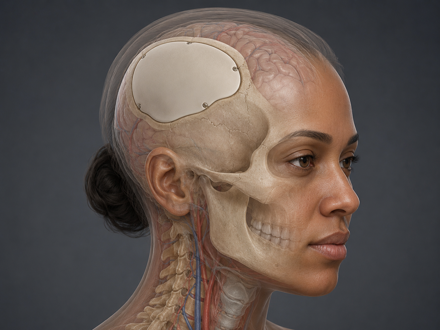
Cranioplasty is surgery to repair or reconstruct a defect in the skull. A skull defect may occur after trauma, prior brain surgery, infection, tumor surgery, or a decompressive craniectomy, which is a procedure where part of the skull is removed to allow swollen brain tissue more room.
Cranioplasty is different from a craniotomy. A craniotomy creates a temporary opening in the skull so the surgeon can access the brain. Cranioplasty reconstructs a skull opening or defect, usually after the original bone has been removed or cannot be replaced safely.
The goal of cranioplasty is to restore protection over the brain, support the scalp, and improve the contour of the skull. De Novo Brain & Spine evaluates adult patients in Stockbridge, Fayetteville, Atlanta, and surrounding communities to help determine whether cranioplasty, continued observation, treatment of an underlying condition, or another surgical plan may be appropriate.
Cranioplasty may be considered when there is a skull defect that needs reconstruction. It does not treat every brain condition by itself, and it does not replace treatment for the original reason the skull defect occurred.
Conditions or situations that may lead to consideration of cranioplasty include:
Cranioplasty is usually considered only when the brain, scalp, wound, infection status, and overall medical condition make reconstruction reasonable.
Cranioplasty may be considered when a patient has an area of missing skull bone that leaves the brain less protected or creates a significant skull contour defect. It may also be considered when the scalp is sunken over the skull defect or when the defect contributes to discomfort, headaches, sensitivity, or difficulty wearing protective devices.
Some patients are evaluated for cranioplasty after a decompressive craniectomy. In that situation, the timing depends on brain swelling, wound healing, infection risk, neurologic status, hydrocephalus, shunt needs, and the patient’s overall recovery.
Cranioplasty may also be considered after infection, tumor surgery, trauma, or prior cranial surgery when the original bone flap could not be replaced or had to be removed. The decision depends on whether reconstruction is safe and whether any active infection, wound problem, or brain swelling has resolved.
Cranioplasty is not performed simply because a prior brain surgery occurred. It is considered when there is a skull defect that requires reconstruction and the expected benefit outweighs the risks.
Doctors determine whether cranioplasty may be appropriate by reviewing the patient’s medical history, neurologic status, imaging, scalp condition, wound healing, infection history, and prior surgical records.
Evaluation may include:
The decision is individualized. A skull defect that looks similar on imaging may require a different plan depending on the patient’s healing, brain condition, infection risk, and overall medical status.
Cranioplasty is performed under anesthesia. The surgeon reopens or works through an incision over the skull defect and carefully lifts the scalp and soft tissue away from the area that needs reconstruction.
The skull defect may be repaired using the patient’s own preserved bone flap, if available and appropriate, or with a manufactured implant. Materials may include titanium, acrylic-based material, synthetic custom implants, mesh, or other reconstructive materials depending on the situation.
The implant or bone flap is shaped and secured to cover the skull defect. The exact material, fixation method, incision plan, and reconstruction approach depend on the size and location of the defect, scalp condition, prior surgery, infection history, and surgeon judgment.
Cranioplasty reconstructs the skull. It is not the same as removing a tumor, evacuating a blood clot, placing a shunt, or treating the underlying cause of a prior brain injury.
The main goals of cranioplasty are to protect the brain, reconstruct the skull defect, support the scalp, and restore a more normal skull contour.
Potential benefits may include improved protection of the brain, improved appearance of the skull contour, improved comfort over the defect, and better ability to participate in rehabilitation or daily activities. In selected patients with sinking skin flap or syndrome of the trephined, cranioplasty may also be associated with improvement in certain neurologic symptoms, but improvement is not guaranteed.
Cranioplasty has important limitations. It does not reverse all brain injury. It does not cure the original condition that led to the skull defect. It does not guarantee improvement in headaches, cognition, strength, speech, balance, or function.
General risks may include infection, bleeding, seizure, swelling, stroke, implant movement, implant exposure, wound healing problems, spinal fluid leak, need for implant removal, bone flap resorption when the patient’s own bone is used, or need for additional surgery. The risks depend on the patient’s prior surgery, scalp condition, infection history, neurologic condition, and overall health.
Treatment planning is individualized. Cranioplasty is one possible step in the larger care plan for a patient with a skull defect.
Alternatives or related planning decisions may include:
The best plan depends on the cause of the skull defect, the patient’s neurologic status, the condition of the scalp and wound, the presence of infection or fluid problems, and the risks and benefits of reconstruction.
Recovery after cranioplasty varies from person to person. It depends on the reason for the skull defect, the size and location of the reconstruction, the implant material, the patient’s neurologic condition, and overall health.
Follow-up usually focuses on incision healing, scalp swelling, neurologic function, headaches, seizure monitoring when relevant, medication use, and imaging when needed. Patients may also continue rehabilitation or other medical care for the original brain injury, stroke, hemorrhage, tumor, or infection.
Patients should follow the surgeon’s instructions about activity, wound care, helmet use if still needed, medications, blood thinners, seizure precautions if applicable, and follow-up appointments. Protection from head injury remains important during recovery.
Seek emergency medical care or call 911 for new seizure, sudden weakness, numbness on one side of the body, trouble speaking, confusion, loss of consciousness, sudden severe headache, or rapid neurologic decline.
After cranioplasty, urgent evaluation is also important for fever, worsening incision redness, drainage, swelling, severe headache, new neurologic symptoms, wound opening, clear fluid leakage, or increasing pain at the surgical site.
Patients with a skull defect who develop new neurologic symptoms, head trauma, wound changes, or signs of infection should seek medical evaluation promptly.
Cranioplasty is used to repair or reconstruct a skull defect. The defect may result from prior brain surgery, decompressive craniectomy, trauma, infection, tumor surgery, or removal of a bone flap.
No. A craniotomy creates an opening in the skull so the surgeon can access the brain. Cranioplasty repairs or reconstructs a skull defect, often after bone has already been removed or cannot be replaced safely.
Cranioplasty may use the patient’s own preserved bone flap or a manufactured implant. Materials may include titanium, acrylic-based material, synthetic custom implants, mesh, or other reconstructive materials depending on the patient’s situation.
Usually not. Cranioplasty reconstructs the skull defect. It does not cure the original condition that led to the skull defect, such as brain injury, stroke, hemorrhage, tumor, infection, or swelling.
Seek urgent medical care for fever, wound drainage, worsening swelling, new seizure, sudden weakness, confusion, severe headache, trouble speaking, loss of consciousness, or rapid neurologic decline.
Schedule a Consultation
Learn if this procedure is right for you.
Endoscopic skull base surgery uses a small camera and specialized instruments to treat selected tumors or defects near the skull base.
Craniotomy for brain tumor resection is brain surgery to remove as much tumor as safely appropriate for diagnosis or treatment.
Shunting of hydrocephalus diverts excess cerebrospinal fluid from the brain to another body area where it can be absorbed.