Meningioma
Evaluation and treatment planning for meningioma, a usually benign tumor of the meninges that may affect brain, skull base, or nerve function.
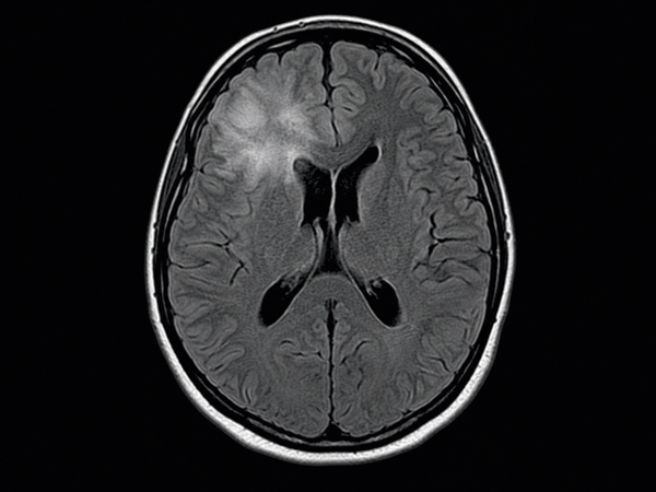
Oligodendroglioma is a type of glioma, which means it is a tumor that begins from glial cells in the brain or spinal cord. Oligodendrogliomas most often occur in the brain and may affect nearby areas that control movement, speech, memory, sensation, vision, mood, or behavior.
In current medical classification, oligodendroglioma is defined by specific molecular findings: an IDH mutation and 1p/19q codeletion. This means that diagnosis depends not only on what the tumor looks like under the microscope, but also on molecular testing. Oligodendrogliomas are generally classified as CNS WHO grade 2 or CNS WHO grade 3 tumors.
De Novo Brain & Spine evaluates adult patients with suspected or confirmed oligodendroglioma to help determine the appropriate next step. Evaluation may include neurological examination, brain MRI, CT imaging, biopsy, image-guided tumor resection, pathology review, molecular testing, or coordination with neuro-oncology and radiation oncology when needed.
Oligodendroglioma symptoms depend on the tumor’s location, size, grade, growth pattern, and swelling around the tumor. Some symptoms develop gradually, while others may appear suddenly.
Common signs and symptoms may include:
Seek emergency medical care or call 911 for a first-time seizure, sudden weakness, sudden speech difficulty, sudden vision loss, severe confusion, loss of consciousness, or a rapidly worsening headache with vomiting or neurological changes.
Most oligodendrogliomas develop without a clearly known cause. A diagnosis of oligodendroglioma does not usually mean that the patient did something to cause the tumor.
Possible risk factors and related medical factors may include:
Doctors use molecular and genetic tumor features to classify oligodendroglioma and guide treatment planning. Key diagnostic features include an IDH mutation and 1p/19q codeletion. Other tumor markers may be reviewed by the care team when appropriate.
These molecular findings are not lifestyle causes. They are tumor features that help confirm the diagnosis, determine tumor classification, and guide treatment decisions.
Oligodendroglioma cannot be diagnosed by symptoms alone. Diagnosis usually requires medical history, neurological examination, brain imaging, tissue diagnosis, and molecular testing.
Common diagnostic steps may include:
The goal of diagnosis is to confirm the tumor type, understand how the oligodendroglioma is affecting the brain, and determine whether observation, surgery, radiation therapy, chemotherapy, or additional specialty care may be needed.
Oligodendroglioma treatment depends on the tumor’s grade, molecular features, size, location, symptoms, imaging findings, neurological examination, surgical risk, and the patient’s overall health. Not every oligodendroglioma requires the same treatment plan.
Treatment options may include:
Surgery may be considered when it can help confirm the diagnosis, reduce tumor burden, relieve pressure on the brain, reduce symptoms, or support the next stage of treatment. The safest plan depends on the oligodendroglioma’s location, grade, molecular findings, and relationship to important brain structures.
Schedule a Consultation
Get an expert opinion about your condition.
Evaluation and treatment planning for meningioma, a usually benign tumor of the meninges that may affect brain, skull base, or nerve function.
Evaluation and treatment planning for brain hemorrhage, including bleeding in or around the brain that may require urgent neurological care.
Evaluation and treatment planning for metastatic brain tumors, also called brain metastases, when cancer spreads to the brain from another site.