Brain Hemorrhage
Evaluation and treatment planning for brain hemorrhage, including bleeding in or around the brain that may require urgent neurological care.

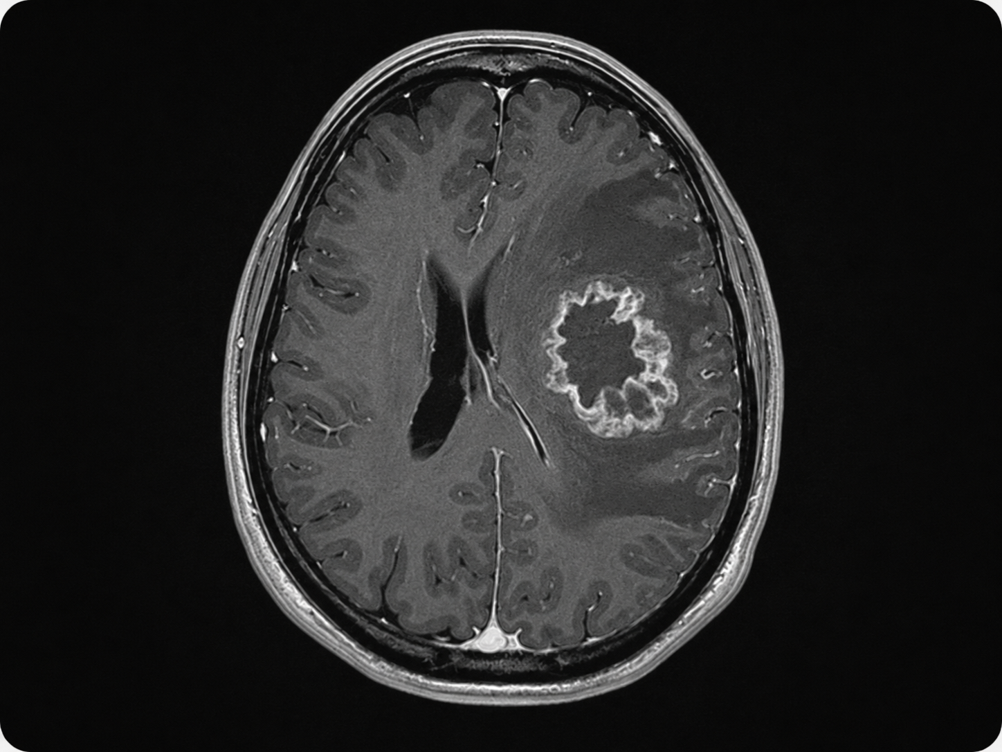
Glioblastoma, also called GBM or glioblastoma multiforme, is an aggressive malignant brain tumor and high-grade glioma. It can grow into nearby brain tissue and may affect areas that control speech, movement, vision, memory, balance, behavior, and personality.
In current medical classification, adult glioblastoma is usually described as glioblastoma, IDH-wildtype, CNS WHO grade 4. This means the diagnosis is based not only on how the tumor looks under the microscope, but also on molecular testing. Important markers may include IDH status, MGMT promoter methylation, EGFR amplification, TERT promoter mutation, and chromosome 7 gain/chromosome 10 loss.
De Novo Brain & Spine evaluates patients with suspected or confirmed glioblastoma using neurological examination, brain MRI, and tissue diagnosis when appropriate. Treatment may involve biopsy, image-guided craniotomy, maximal safe brain tumor resection, radiation therapy, temozolomide chemotherapy, tumor treating fields, clinical trials, and coordinated neuro-oncology care.
Glioblastoma symptoms depend on the tumor’s size, location, growth pattern, and swelling around the tumor. Symptoms may develop gradually or appear suddenly.
Common signs and symptoms may include:
Seek emergency medical care or call 911 for a first-time seizure, sudden weakness, sudden speech difficulty, severe confusion, loss of consciousness, or a rapidly worsening headache with vomiting or neurological changes.
Most glioblastomas are sporadic, meaning they develop without a clear inherited cause. In many patients, there is no known preventable reason why the tumor formed.
Possible risk factors may include:
Doctors may also review molecular and genetic tumor features to better classify the glioblastoma and guide treatment planning. These may include IDH-wildtype status, MGMT promoter methylation, EGFR amplification, TERT promoter mutation, and chromosome 7 gain/chromosome 10 loss.
Glioblastoma is not usually caused by a head injury, routine daily activity, or a single lifestyle factor. Diagnosis and treatment decisions depend on imaging, pathology, molecular testing, neurological function, and the patient’s overall health.
Glioblastoma cannot be diagnosed by symptoms alone. Diagnosis usually requires a combination of medical history, neurological examination, brain imaging, and tissue testing.
Common diagnostic steps may include:
These results help the care team understand the tumor type, confirm the diagnosis, plan treatment, and determine whether surgery, radiation, chemotherapy, clinical trials, or other therapies may be appropriate.
Glioblastoma treatment is individualized. The right plan depends on the tumor’s size, location, molecular profile, symptoms, neurological examination, surgical risk, age, overall health, and goals of care.
Treatment options may include:
Surgery is not appropriate for every patient with glioblastoma. When surgery is considered, the goal is usually to obtain a diagnosis, reduce tumor burden, relieve pressure on the brain, and preserve neurological function as safely as possible.
Schedule a Consultation
Get an expert opinion about your condition.
Evaluation and treatment planning for brain hemorrhage, including bleeding in or around the brain that may require urgent neurological care.
Evaluation and treatment planning for metastatic brain tumors, also called brain metastases, when cancer spreads to the brain from another site.
Evaluation and treatment planning for brain aneurysms, also called cerebral or intracranial aneurysms, including unruptured and ruptured aneurysms.